The results pointed out no statistically significant differences of micronucleated oral mucosa cells before versus after X-ray exposure for both oral sites of smokers or to non-smokers. compared and evaluated DNA damage (micronucleus) and cellular death (pyknosis, karyolysis, and karyorrhexis) in exfoliated oral mucosa cells from smokers and nonsmokers submitted to dental X-ray using two anatomic sites: buccal mucosa and lateral border of the tongue. In our study, the difference of micronucleus and other nuclear alterations in buccal mucosa was 0.087 ± 2.0 and 11.38 ± 2.49, and gingiva was 0.013 ± 0.13 and 5.38 ± 1.89, respectively, showed statistically significant results ( P 0.05).Īngelieri et al.
Kv^5 6 exposure x rays full#
The results in the present study are consistent with other studies conducted on extraoral projections, but in the present genotoxic and cytotoxic changes were seen in patients exposed to full mouth set of radiographs, which is not reported in the literature to best of our knowledge. The study concluded that the complete set of radiographs requested in the orthodontic planning may not be a factor that induces chromosomal damage, however, it is able to promote cytotoxicity. evaluated DNA damage (micronucleus (MN)) and cellular death (pyknosis, karyolysis, and karyorrhexis) in exfoliated buccal mucosa cells from children undergoing orthodontic radiographs (lateral cephalographic, posteroanterior view, panoramic, full periapical exam, and bitewing).

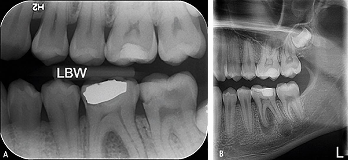
Taken together, these results indicated that panoramic dental radiography might not induce chromosomal damage but may be cytotoxic. However, on the other hand, radiation did cause other nuclear alterations closely related to cytotoxicity including karyorrhexis, pyknosis, and karyolysis. There was a significant increase ( P 0.05), whereas the condensation of the chromatin and the karyorrhexis increased significantly after exposure ( P 0.05) increase in the frequency of cells with micronuclei. Comparison of difference of micronuclei between buccal mucosa and gingiva of full mouth exposure was found to be significant ( P 0.05). The mean of other nuclear alterations indicating cytotoxicity after exposure to full mouth radiographs was significant ( P 0.05), and. Results: The mean micronuclei frequency in buccal mucosa and gingiva of the group was increased after exposure but the difference was not significant statistically ( P > 0.05). The samples were stained with the paponicolaou method and accessed for micronuclei and karyolysis, pyknosis, and karyorrhexis. Materials and Methods: Cytological smears were taken from the buccal mucosa and gingiva of the study group just before exposure to X-rays and 10 days after exposure to X-rays. Study Design: The study group consisted of 30 patients exposed to conventional full mouth radiographs.
Kv^5 6 exposure x rays series#
Objectives: To evaluate and compare mutagenicity (micronucleus) and cytotoxicity (karyorrhexis, pyknosis, and karyolysis) in exfoliated buccal mucosa cells of patients following conventional full mouth series of radiographs.


Therefore, outlining the cytogenetic effects induced by X-rays is necessary to identify the degree of cancer risk and minimize potential risks to patients and clinicians.

However, it is well-known that ionizing radiation causes damage (including single- and double-strand breaks) to DNA and DNA–protein crosslinks and induces cellular death. Background: In the past decades, X-rays have been used widely for diagnosis in dentistry.


 0 kommentar(er)
